

Zubal Digital Phantoms and Their Applications in dosimtery + download files
Comprehensive Article: Zubal Digital Phantoms and Their Applications in Nuclear Medicine
Abstract
Zubal digital phantoms are among the most authoritative digital anatomical models in nuclear medicine and radiotherapy research. These phantoms are based on real human imaging data and provide high-precision differentiation of various tissues and organs. This article provides a comprehensive introduction to the different types of Zubal phantoms, their technical specifications, dimensions, and extensive applications across various domains of radiation medicine.
Introduction
In the era of digital medical imaging and precise radiation therapies, there is a critical need for standardized anatomical models for calibration, evaluation, and optimization of imaging and treatment systems. Zubal digital phantoms, developed in the early 1990s by the research team at Yale University, represent a scientific response to this fundamental need. These phantoms have found widespread applications not only in academic research but also in industry and clinical settings.
History and Development
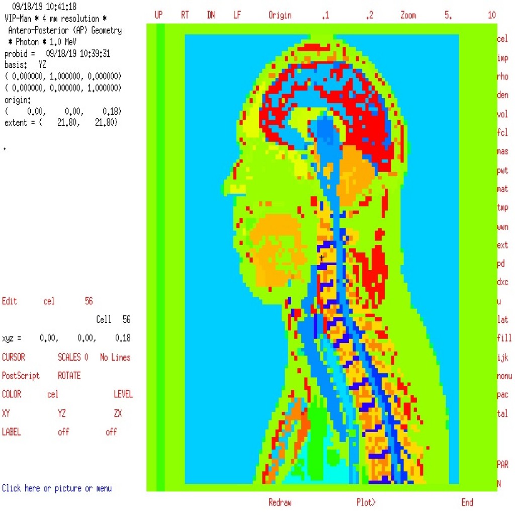
The Zubal phantom was first introduced in 1994 by George Zubal and his colleagues at Yale University. This phantom was created based on high-resolution MRI images from a healthy male volunteer. The differentiation of various tissues and organs was performed manually by radiology specialists, resulting in a highly precise anatomical model with 125 distinct regions.
Types of Zubal Phantoms
1. Head Phantom (MRI Head Phantom)
Designed specifically for brain and skull studies, this phantom offers 256×256 pixel resolution with 128 slices, providing detailed insights into brain structures.
2. Whole Body Phantom (Zubal Whole Body Phantom)
The most famous and widely used phantom in this collection, covering the entire body from head to toes. This phantom serves as the foundation for most dosimetry and optimization studies in nuclear medicine.
3. Arms Folded Phantom
Designed to simulate real scanning conditions in PET/CT systems where patients typically place their arms above their head or across their chest.
4. Arms Down Phantom
Another model for simulating different patient positions during imaging, useful for comparative studies and evaluating the impact of positioning on image quality.
Technical Specifications Table
| Phantom Name | Filename | Width (pixels) | Height (pixels) | Number of Slices | Voxel Size (mm³) |
|---|---|---|---|---|---|
| Whole Body Phantom | voxel_man.dat | 128 | 128 | 243 | 4.17×4.17×4.17 |
| Head Phantom | det_head_u2_med.dat | 256 | 256 | 128 | 1.0×1.0×1.0 |
| Arms Folded | MAN_TISSUES_3-6.dat | 493 | 87 | 147 | Variable |
| Arms Down | vox_tiss8.dat | 192 | 96 | 498 | Variable |
Organ Identification System
The Zubal phantom uses a unique numerical system to identify each tissue or organ. This system includes 125 different identifiers, with the most important being:
Vital Organs and Their Identifiers:
-
0: Outside phantom (background)
-
1: Skin
-
2: Brain
-
11: Heart
-
12: Liver
-
14: Kidney
-
23: Blood pool
-
26: Bone marrow
-
40: Bladder
Neural Structures:
-
77: Cerebellum
-
83: White matter
-
109: Thalamus
-
111: Corpus callosum
-
113: Cerebral falx
Bone Structures:
-
4: Skull
-
5: Spine
-
6: Rib cage & sternum
-
7: Pelvis
-
125: Teeth
Applications of Zubal Phantoms
1. Radiation Dosimetry Calculations
-
Determining dose distribution in radiotherapy
-
Evaluating organ doses in diagnostic imaging
-
Radiation protection studies for staff and patients
2. Imaging Protocol Optimization
-
Optimizing CT scan parameters
-
Developing new PET and SPECT protocols
-
Assessing the impact of imaging parameters on patient dose
3. Algorithm Development and Evaluation
-
Testing image reconstruction algorithms
-
Evaluating noise reduction methods
-
Developing artifact correction techniques
4. Education and Standardization
-
Educational tool for medical physics students
-
Reference standard for comparing different systems
-
Foundation for developing new phantoms
5. Monte Carlo Simulations
-
Use in GATE, Geant4, and FLUKA software
-
Radiation transport studies in different tissues
-
Development of new physical models
Advantages of Zubal Phantoms
1. High Anatomical Accuracy
-
Based on real imaging data
-
Precise anatomical boundary differentiation
-
Realistic representation of spatial relationships between organs
2. Comprehensiveness
-
Full body coverage from head to toe
-
Inclusion of all vital organs
-
Differentiation of soft and hard tissues
3. Accessibility
-
Available to the global scientific community
-
Simple and interpretable format
-
Complete documentation and user guides
4. Standard Compliance
-
Compatible with DICOM standards
-
Usable in most medical software
-
Foundation for international standards
Limitations and Challenges
1. Inherent Limitations
-
Based on anatomy of a single individual
-
Lack of population diversity
-
Absence of pathological conditions
2. Technical Limitations
-
Fixed spatial resolution
-
Lack of dynamic variability (breathing, heartbeat)
-
Absence of histological microstructure information
3. Evolutionary Needs
-
Need for updates with new technologies
-
Development of female and pediatric versions
-
Addition of pathological tissues
Future of Digital Phantoms
1. Next-Generation Phantoms
-
Population-based phantoms
-
Dynamic and 4D models
-
Molecular and functional phantoms
2. Integration with Artificial Intelligence
-
Personalized phantom generation
-
Tissue response prediction to radiation
-
Automated protocol optimization
3. Expansion of Applications
-
Use in heavy particle radiotherapy
-
Applications in therapeutic nuclear medicine
-
Use in developing new radiopharmaceuticals
Conclusion
Zubal digital phantoms, as one of the most fundamental research tools in medical physics and nuclear engineering, have played a vital role in advancing radiation sciences. Their simple structure, anatomical accuracy, and comprehensiveness have made them a gold standard in both academic and industrial research. Despite existing limitations, the Zubal phantom remains a valuable starting point for developing more complex and personalized models.
Continuous development of these phantoms and their integration with emerging technologies such as artificial intelligence and advanced modeling will open new horizons in radiation medicine research and contribute to more precise and safer healthcare delivery.
References and Useful Links
Scientific Resources
-
Zubal, G. et al. (1994) - Foundational paper introducing the phantom
-
Specialized journals: Medical Physics and Physics in Medicine & Biology
-
International medical physics conferences
Data Access
-
Official Yale University website
-
International scientific data repositories
-
Zubal phantom user community
Scientific Communities
-
American Association of Physicists in Medicine (AAPM)
-
European Federation of Organizations for Medical Physics (EFOMP)
-
International research networks
This article serves as a comprehensive reference for researchers, students, and professionals working in nuclear medicine, radiotherapy, medical physics, and nuclear engineering who wish to utilize Zubal phantoms in their research and projects.